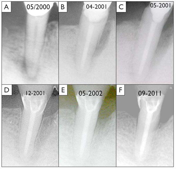
Fig. (1) Radiographic sequence of the presented case: A) baseline image of tooth 45 showing the periapical translucency in May 2000, B)
situation after the endodontic therapy and the development of a 9 mm pocket (April 2001), C) radiograph after periodontal surgery and ablative
removal of the cemental tear (May 2001), D) immediately after restorative treatment (December 2001), E) situation one year after surgery
and 6 month after oral rehabilitation (May 2002) and finally F) status after more than ten years after the surgical intervention (September
2011).