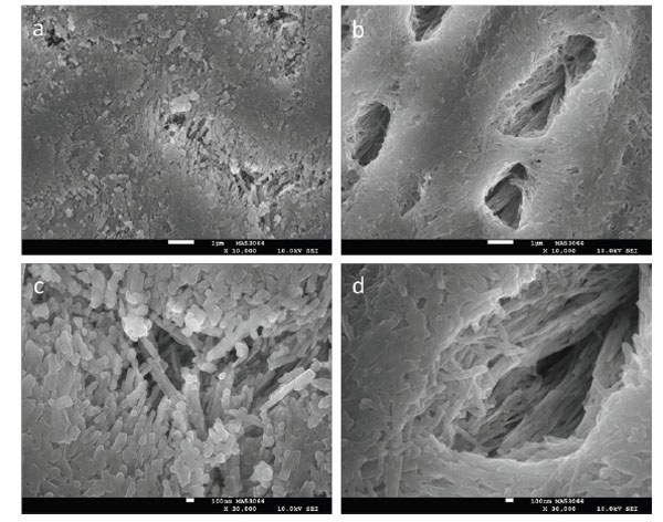
Fig. (1)
SEM images at 10,000x (
a
,
b
) and 30,000x (
c
,
d
) of enamel initially eroded for 30 minutes with 1% citric acid (
a
,
c
) or 120 minutes with 0.3% citric acid (
b
,
d
). The white scale bars correspond to 1 μm (
a
,
b
) and 100 nm (
c
,
d
).