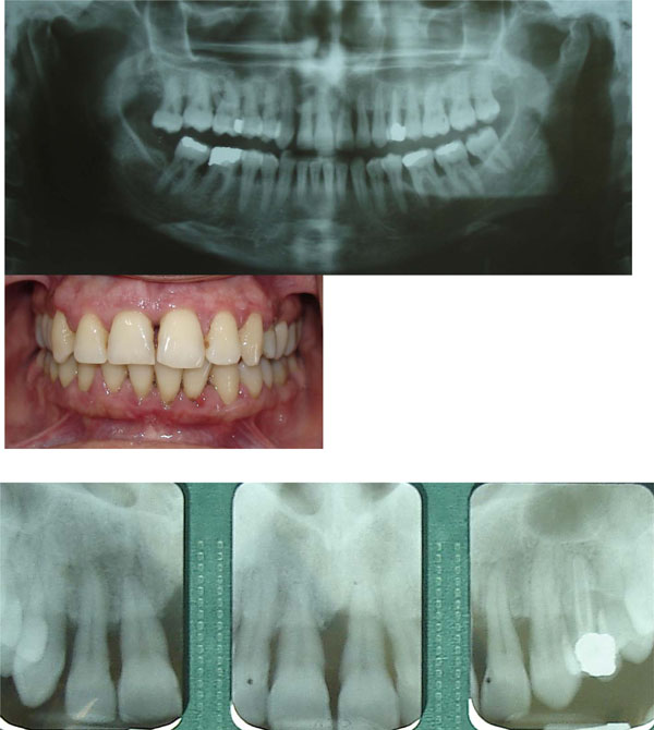
Fig. (1)
Above:
Initial panoramic and clinical views of occlusion and tooth alignment.
Below:
Periapical radiographs of the maxillary incisors.