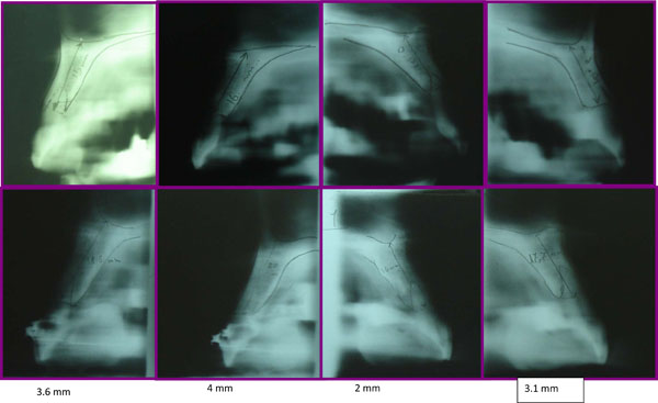
Fig. (2)
Tomographic scans of the maxillary incisors before treatment (
top row
) and after treatment (
bottom row
).
Left to right:
right lateral incisor, right central incisor, left central incisor, left lateral incisor.