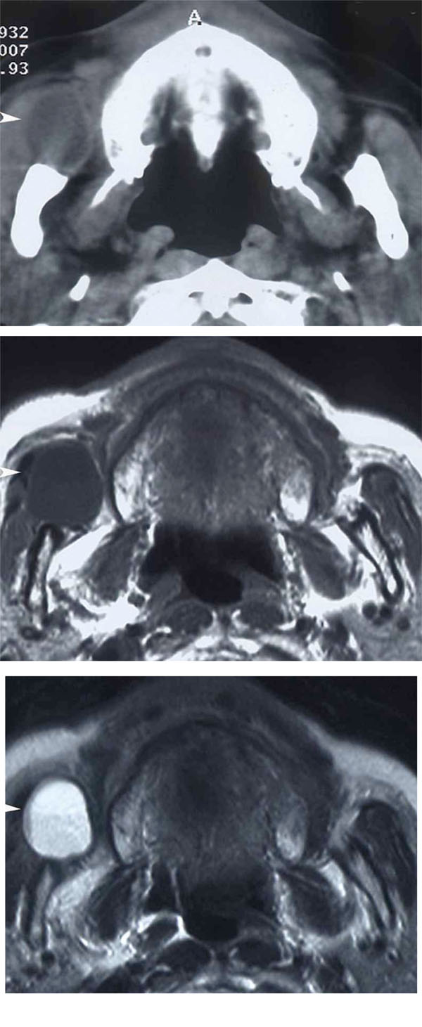
Fig. (2) Imaginga: Axial CT, b: Axial MRI (T1WI), c: Axial MRI (T2WI)A well-circumscribed low density mass is observed in the right buccal region anterior to the masseter muscle in CT (arrow head). It shows a low signal on T1-weighted MRI and a heterogeneous high signal on T2-weighted MRI (arrow head).