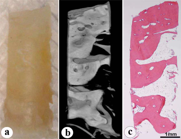
Fig. (2)
Alveolar bone findings.
a
: Appearance of the alveolar bone sample.
b
: 3D-microCT image of the longitudinal section, through the center of the sample.
c
: Hematoxylin-eosin staining of the same section as in (b).