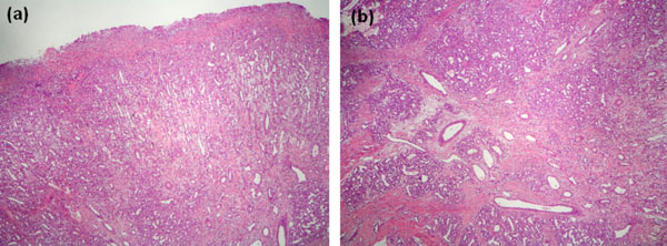
Fig. (4a) Microscopic appearance. Under an ulcerated surface there is an extensive granulation tissue reaction characterized by abundant small size vessels arranged perpendicularly to surface. At the bottom of the image, the characteristic appearance of pyogenic granuloma can be seen on the right (H&E, original magnification 10x).
(b). At a higher power, the typical histologic features of pyogenic granuloma consist in a proliferation of capillary sized vessels arranged surrounding larger vessel structures producing its typical lobular distribution. There is stromal fibrosis dissecting between the capillary lobules. (H&E, original magnification 20x).

