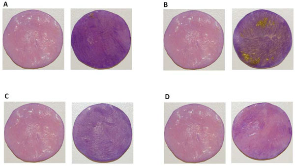
Fig. (1) Biofilm formation on DBR disc surfaces. Representative images after crystal violet staining of biofilms formed on DBR discs by F.
nucleatum ATCC 23726 (A) T. forsythia ATCC 43037 (B) V. atypica PK 1910 (C) K. pneumoniae IA 565 (D) For each set, the image on the
left was a control DBR disc without biofilms growing on the surface, the image on the right was a DBR disc with biofilms growing on the
surface. The experiment was performed in triplicate.

