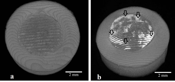
Fig. (5) (a) 3D Micro-CT images of the unprocessed sample which the outer side is restorative material and the inner side is pulp capping
material and (b) 3D Micro-CT images of the sample which exposed to copper. (image has white intense regions shown by the black arrow
due to Cu treatment on the surface).