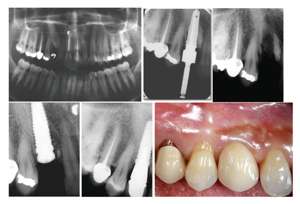
Fig. (1) Patient # 1. a) Panoramic radiograph with the unerupted canine. b) Radiograph showing the drilling through the impacted canine. c)
Radiograph after removing the drill. Reduction in radio-opacity is related to the removal of dental tissue. d) Implant placement after removing
the mobile fragment of the crown on the mesial side. e) Periapical radiograph at the 8-year control. f) Vestibular clinical view at the 8-year
control. The papilla length is similar to the adjacent natural teeth.