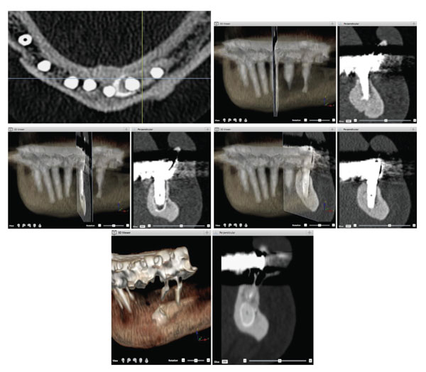
Fig. (2) Patient # 2. a) Preoperative tomodensitometric examination displaying a 3D reconstruction and a transverse section at the impacted
ectopic horizontal premolar. The impacted tooth is in the middle of the mandible just beneath the root of the failing teeth. b) Localisation of
implant WHO # 32 (ADA # 23) and corresponding transverse section of the tomodensitometric examination at the 8-year control. c) Localisation
of implant WHO # 33 (ADA # 22) and corresponding transverse section at the 8-year control. d) Localisation of implant WHO # 34
(ADA # 21) and corresponding transverse section at the 8-year control. e) Axial section of the tomodensitometric examination at the 8-year
follow-up. Note the distinct position of each implant in contact with the various parts of the impacted premolar, the cuspid of the crown, the
crown and the root. No specific deleterious radiographic feature could be observed.