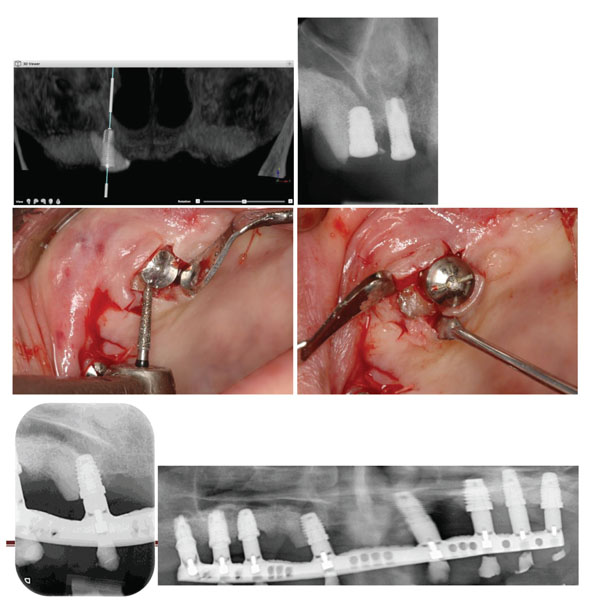
Fig. (3) Patient # 3. a) Pre-operative panoramic radiograph with the radicular part of the canine. b) Post-operative radiograph of the implant
encroaching the root. c) Flattening the distal part of the root of the impacted canine to accommodate the abutment. d) Flattened distal part of
the root before suturing over the abutment. e) Periapical radiograph of the implant incroaching upon the canine at the 5-year control. f) Panoramic
radiograph of the implant-supported prosthesis at the 5-year control.