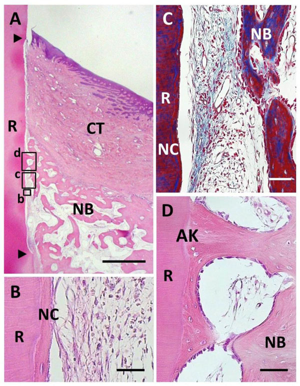
Fig. (4) Histological findings in BMP group. (A) Alveolar bone formation occurred along the root surface. Coronal and apical notches are indi-cated by arrowheads. (B) Higher magnification of framed area (b) in (A). New cellular cementum was detected on the BMP-treated root surface. (C) Higher magnification of framed area (c) in (A). Little periodontal ligament formation was observed between new bone and cementum. (D) Higher magnification of framed area (d) in (A). An-kylosis was frequently noted. R, root; CT, gingival connective tissue; NB, new bone; NC, new cementum; AK, ankylosis. Staining: hema-toxylin and eosin (A, B, D) and Masson’s trichrome (C). Scale bars: (A) = 1 mm; (B) = 25 µm; (C and D) = 50 µm.