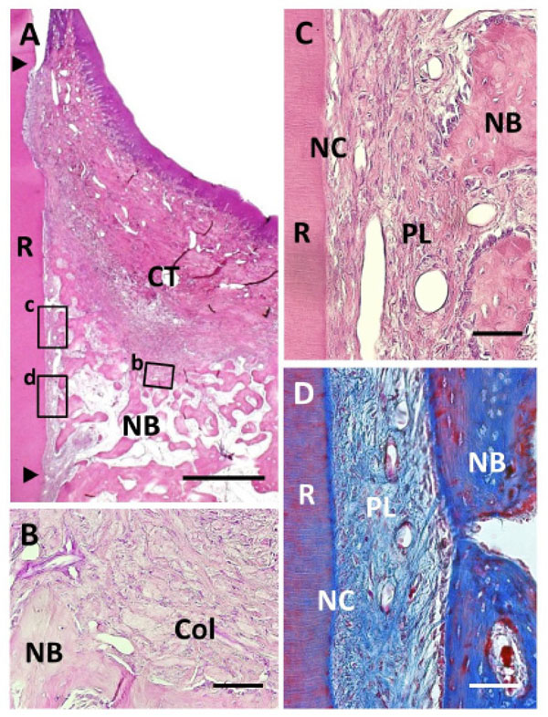
Fig. (5) Histological findings in Col group. (A) Formation of new bone was observed in the defect. Coronal and apical notches are indicated by arrowheads. (B) Higher magnification of framed area (b) in (A). Cell ingrowth was observed in the residual collagen hydrogel scaffold. (C) Higher magnification of the framed area (c) in (A). Thin layer of ce-mentum-like tissue was observed on the root dentin surface. (D) High-er magnification of the framed area (d) in (A). Periodontal ligament tissue with functionally oriented fibers was reestablished between the new bone and new cementum-like tissue. R, root; CT, gingival connec-tive tissue; NB, new bone; NC, new cementum; Col, Collagen hydro-gel scaffold; PL, periodontal ligament. Staining: hematoxylin and eosin (A, B, C) and Masson’s trichrome (D). Scale bars: (A) = 1 mm; (B, C and D) = 50 µm.