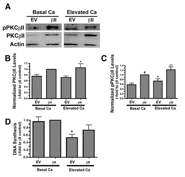
Fig. (3) Overexpressed PKCβII is autophosphorylated/activated in response to an elevation of extracellular calcium concentration in mouse keratinocytes but has no effect on calcium-inhibited proliferation. Mouse keratinocytes were nucleofected with wild-type PKCβII (βII) or empty vector (EV) and then cultured in the presence of basal (25µM) or a moderately elevated extracellular calcium level (Ca; 125µM) for 24h. Cells were harvested and lysates were resolved on 8% SDS gels, transferred to PVDF membranes and probed with antibodies recognizing total PKCβII, autophosphorylated PKCβII and actin. (A) A representative experiment is illustrated. (B) Total PKCβII and (C) pPKCβII levels were quantified, normalized to actin and expressed relative to the PKCβII-transfected cells under basal conditions. Data represent the means ± SEM from at least 3 separate experiments. For total PKCβII, *p<0.05 vs EV or EV+Ca and for autophosphorylated PKCβII, #p<0.01 vs EV, *p<0.01 vs EV+Ca, ^p<0.001 vs EV, +p<0.05 vs βII. (D)Keratinocytes nucleofected with wild-type PKCβII (βII) or empty vector (EV) were cultured in the presence of basal (25µM) or a moderately elevated extracellular calcium level (Ca; 125µM) for 24h. [3H]Thymidine was added to the medium for 1 hour and DNA synthesis measured as described in Materials and Methods. [3H]Thymidine incorporation into DNA is expressed as the percentage of the control value and shown as the mean ± SEM (n=4; *p<0.01 vs EV or βII).