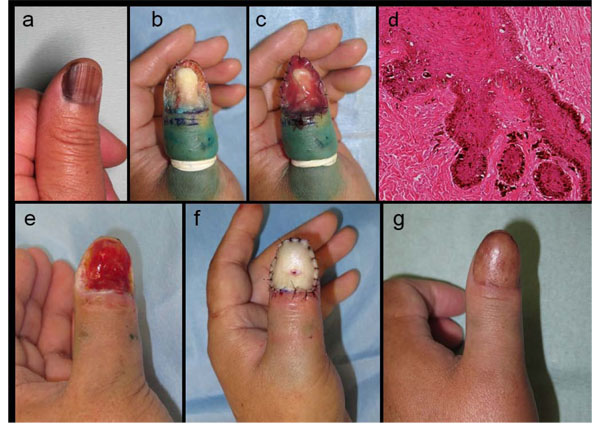
Fig. (1)
Clinical course and histopathological features of case 1.(a) Left thumb NUM in situ. (b) Wide skin excision extending vertically to the subperiosteal layer. (c) INTEGRA® was directly placed onto the exposed bone and tissue. (d) Histologically, numerous atypical melanocytes were scattered along the basement membrane. (e) The regenerated dermal tissue induced by INTEGRA® fully covered the defect within one month after the lesional excision as the first phase operation. (f) Reconstruction with a PSVNSG technique using a graft from the patient’s clavicular skin. (g) Postoperative appearance one year after the skin grafting.