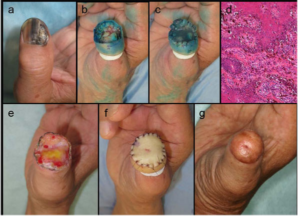
Fig. (3)
Clinical course and histopathological features of case 3.(a) NUM with nail destruction on the right thumb. (b) Wide skin excision extending vertically to the subperiosteal layer and amputation of the distal phalanx. (c) INTEGRA® was sutured directly onto the surgical defect without debriding the bone. (d) Atypical melanocytes were seen along the basement membrane with nodular invasion at the nail bed. (e) Regenerated dermal tissue induced by INTEGRA® fully covered the defect within one month after the first phase operation. (f) Reconstruction with PSVNSG utilizing a graft from the patient’s clavicular skin. (g) Postoperative appearance at one year after the skin grafting.