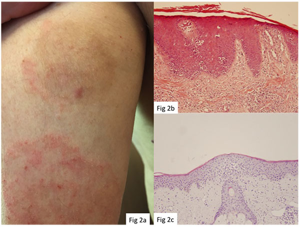
Fig. (2) a. Hyperpigmented postinflammatory target MF lesion (yellow arrow) and erythemato-squamous control MF lesion (red arrow) 2 months after treatment. 2b. Histology illustrating patch/plaque stage MF before and 2c. Histological clearing 2 months after treatment (H/E histochemical stain, x10).