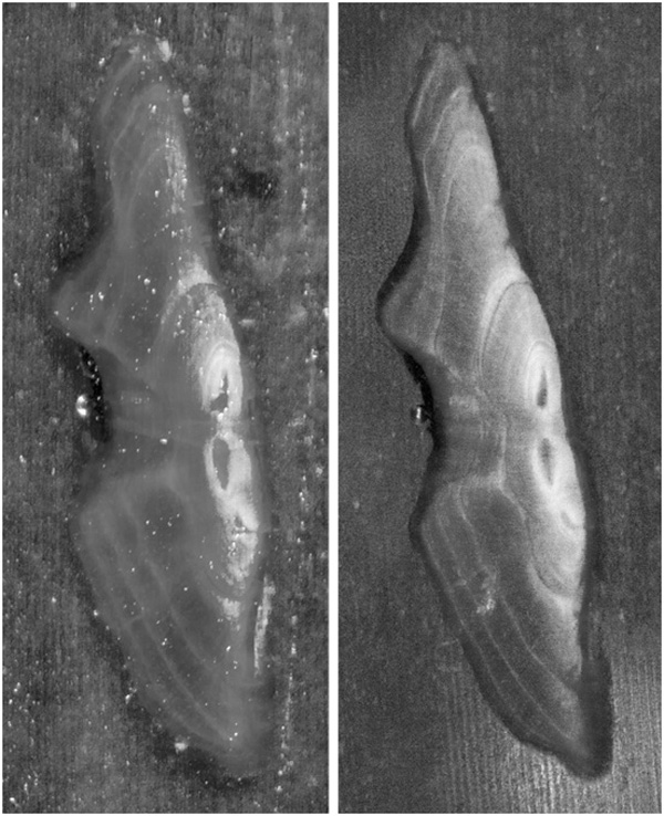
Fig. (2) Digital images of a single walleye otolith section viewed using an alternative method (left) and the otolith illumination device (right). Note the increased clarity of annuli on the otolith section viewed using the otolith illumination device.