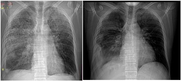
Fig. (4)
Case 1, Radiograph on the left showed cavity peripheric nodules; two on the left before the treatment bilateral pulmonary consolidations and pleural and two on the right during the treatment effusion (before the treatment). On the right there is right pulmonary consolidation (during the treatment).