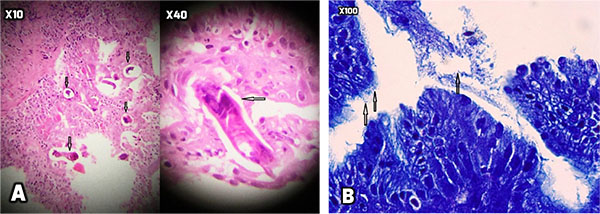
Fig. (3)
Gastric mucosa showed focal erosion, surface irregularity with mixed inflammatory cells infiltrated in lamina propria. Also, some S. stercoralis larvae (A) were found in gastric pits with the mild colonization of H. Pylori (B). The figures shown in increases of 10, 40 and 100 times.