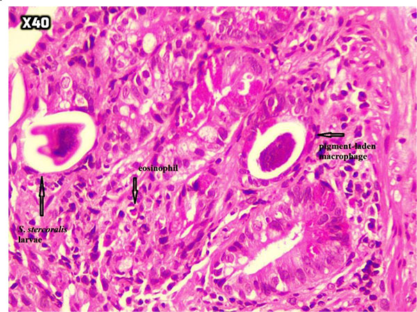
Fig. (4)
Ulceration and granulation tissue formation with focal crypt distortion, cryptitis, and focal crypt abscess formation with increase in inflammatory cells rich in eosinophil and some pigment-laden macrophages as well as some parasite-like structures (H&E x40).