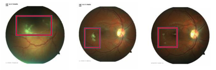
Fig. (1)
The above images track the improvement of the patient on fundoscopic examination beginning in April 2016 through August 2016. A large pre-retinal abscess can be noted on the first image as well as irregular vessel structure. Improvement of the abscess and vessel structure can be observed in the subsequent pictures.