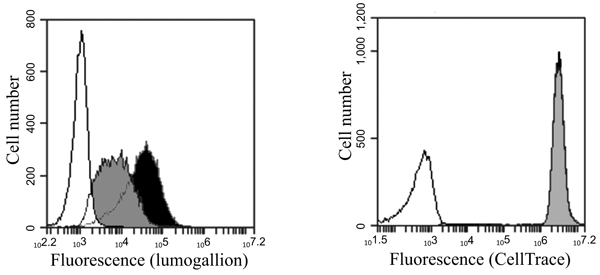
Fig. (6A) Flow cytometry of cells stained with CellTrace™ Far Red immediately after incubation with aluminium adjuvants. THP-1 cells stained with the cytoplasm stain CellTrace™ Far Red were incubated with lumogallion labelled ABAs. The cells were separated from free adjuvant particles and re-suspended in culture medium. Experimental conditions as described in the Materials and Methods section. Left histogram: Aluminium adjuvant fluorescence intensity (lumogallion) associated with THP-1 cells immediately after separation from free ABAs in the culture medium. Black histogram: Cells incubated with lumogallion labelled Alhydrogel®. Grey histogram: Cells incubated with lumogallion labelled Adju-Phos®. White histogram: Cells incubated in culture medium. Right histogram: Cytoplasm fluorescence intensity (CellTrace™ Far Red) from THP-1 cells immediately after separation from free ABAs in the culture medium. Black histogram: Cells incubated with lumogallion labelled Alhydrogel®. Grey histogram: Cells incubated with lumogallion labelled Adju-Phos®. White histogram: Non-stained cells in culture medium. Histograms of cells stained with CellTrace™ Far Red and incubated with the two forms of lumogallion labelled ABAs showed the same fluorescence intensity (black and grey histograms are completely overlapping).