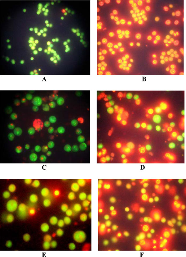
Fig. (2) Fluorescence microscopic images of untreated control U937 (A), K562 (C) and HL-60 (E) and LCLE treated U937 (B), K562 (D)
and HL-60 (F) cells. The control cells were with intact nuclei and gave bright green fluorescence whereas treated cells showed intense orange-
red fluorescence showing signs of apoptosis.