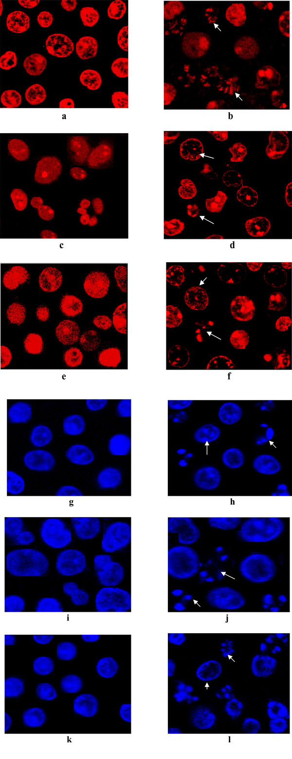
Fig. (3) Confocal microscopic images of untreated control U937 (A, G), K562 (C, I), HL-60 (E, K) and LCLE treated U937 (B, H), K562
(D, J) HL-60 (F, L) cells. The control cells were with intact nuclei whereas LCLE treated cells indicated apoptotic changes like nuclear disintegration
and formation of apoptotic bodies, shown by the arrowheads. A to F are cells stained with propidium iodide and G to L are cells
stained with Hoescht 33342.