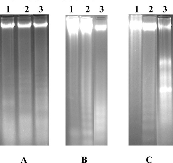
Fig. (5) The gel pattern of DNA samples isolated from (A) untreated
control U937 cells (lane 1), U937 cells treated with 100
µg/ml of LCLE (lane 2), U937 cells treated with 200 µg/ml of standard
anti-cancer drug, AraC (lane 3), (B) untreated control K562
cells (lane 1), K562 cells treated with 100 µg/ml of LCLE (lane 2),
K562 cells treated with 200 µg/ml of standard anti-cancer drug,
AraC (lane 3), and (C) untreated control HL-60 cells (lane 1), HL-
60 cells treated with 200 µg/ml of standard anti-cancer drug, AraC
(lane 2) and HL-60 cells treated with 100 µg/ml of LCLE (lane 3).
Treatment with both the standard anti-cancer drug, AraC and LCLE
showed distinct DNA ladder formation indicating the process of
apoptosis in all the three human leukemic cell lines.