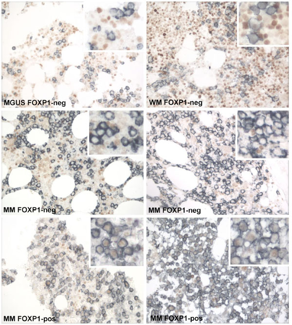
Fig. (5) Double immunoenzymatic labelling of bone marrow samples; FOXP1 brown and CD138 blue. As indicated by the top four panels
the majority of pre-malignant and malignant CD138+ plasma cells lacked expression of the FOXP1 protein, although there were many
CD138-FOXP1+ cells in this tissue. The detection of >30% FOXP1+/CD138+ cells was restricted to two MM cases, one from Oxford (bottom
left) and the other from Zagreb (bottom right).