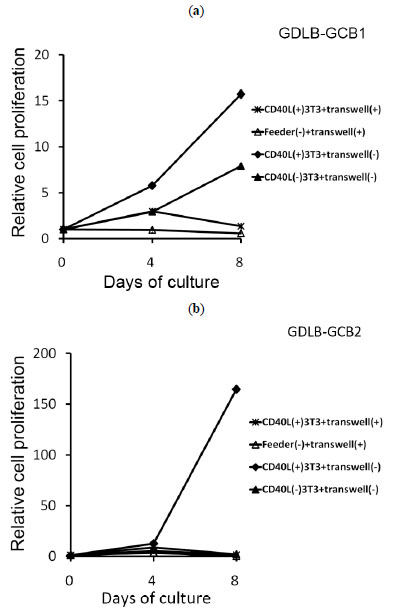
Fig. (5) Growth curves of the established cell lines (GDLBGCB1
and GDLB-GCB2) without direct contact with feeder
cells. When tumor cells and CD40L+3T3 cells were separated with
a micropore membrane, the proliferation was inhibited (ж). The
positive control consisted of three conditions: culture with the inner
cup without feeder cells (Δ), culture without the inner cup with
CD40L+3T3 cells (♦), and culture without the inner cup with
CD40L-3T3 cells (▲). The cells of GDLB-GCB1 stimulated with
CD40L-3T3 showed cell growth on day 8 (Fig. 5a). However, when
the cell lines were cultured with CD40L-3T3 cells in a 96-well
plate, the both cell lines stopped growing as expected. Each
condition was repeated in duplicate, and the cell number was
counted two times in each well on days 4 and 8. Data are presented
as the mean ± SE. Abbreviation: CD40L, CD40 ligand;
CD40L+3T3, NIH Swiss mouse embryonic fibroblast cell line
transfected with CD40L; CD40L-3T3, NIH Swiss mouse embryonic
fibroblast cell line untransfected with CD40L.