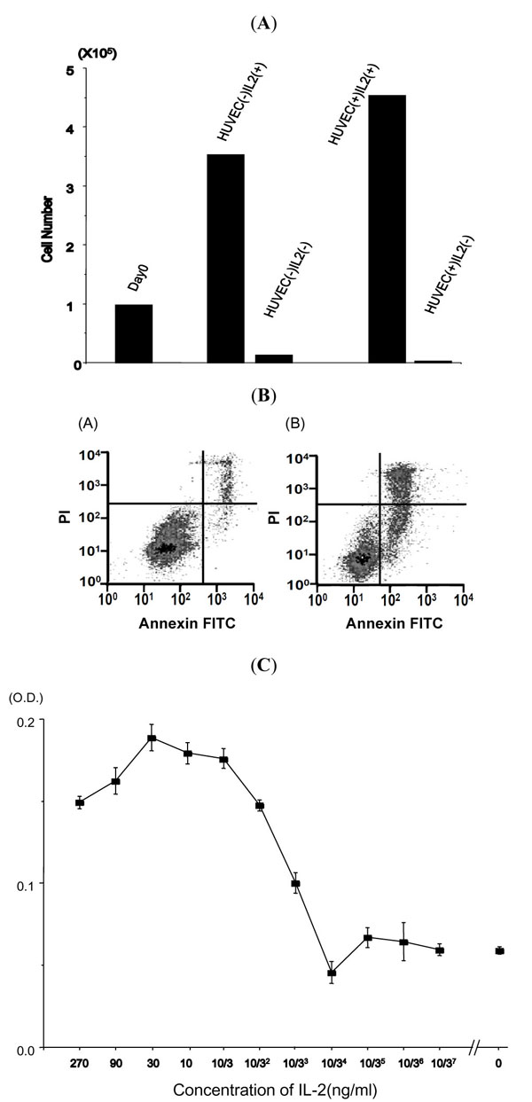
Fig. (3) Promotion and inhibition of HU-ATTAK cell growth by IL-2. (A) HU-ATTAK cell death without IL-2. HUVEC was cultured in a 12-well cluster dish on day -1. 105 cells of HUATTAK were cultured with or without HUVEC or IL-2. The numbers of viable cells were counted on day 3. This is a representative of two independent experiments. (B) Apoptotic cell death of HU-ATTAK in the absence of IL-2. Flow cytometric analysis of apoptosis after 48 hours from the deprivation of IL-2 is shown. The fluorescence intensities of AN (abscissa) and PI (vertical) are plotted. A representative of three experiments is shown. (C) Dose response curve of various concentrations of IL-2 in the presence of HUVEC. HUVECs were cultured in a 12-well cluster dish on day -1. 105 cells of HU-ATTAK were cultured in 2 ml of medium containing serially diluted IL-2. The mean value of quadruplicate assays is shown. This is a representative of two independent experiments.