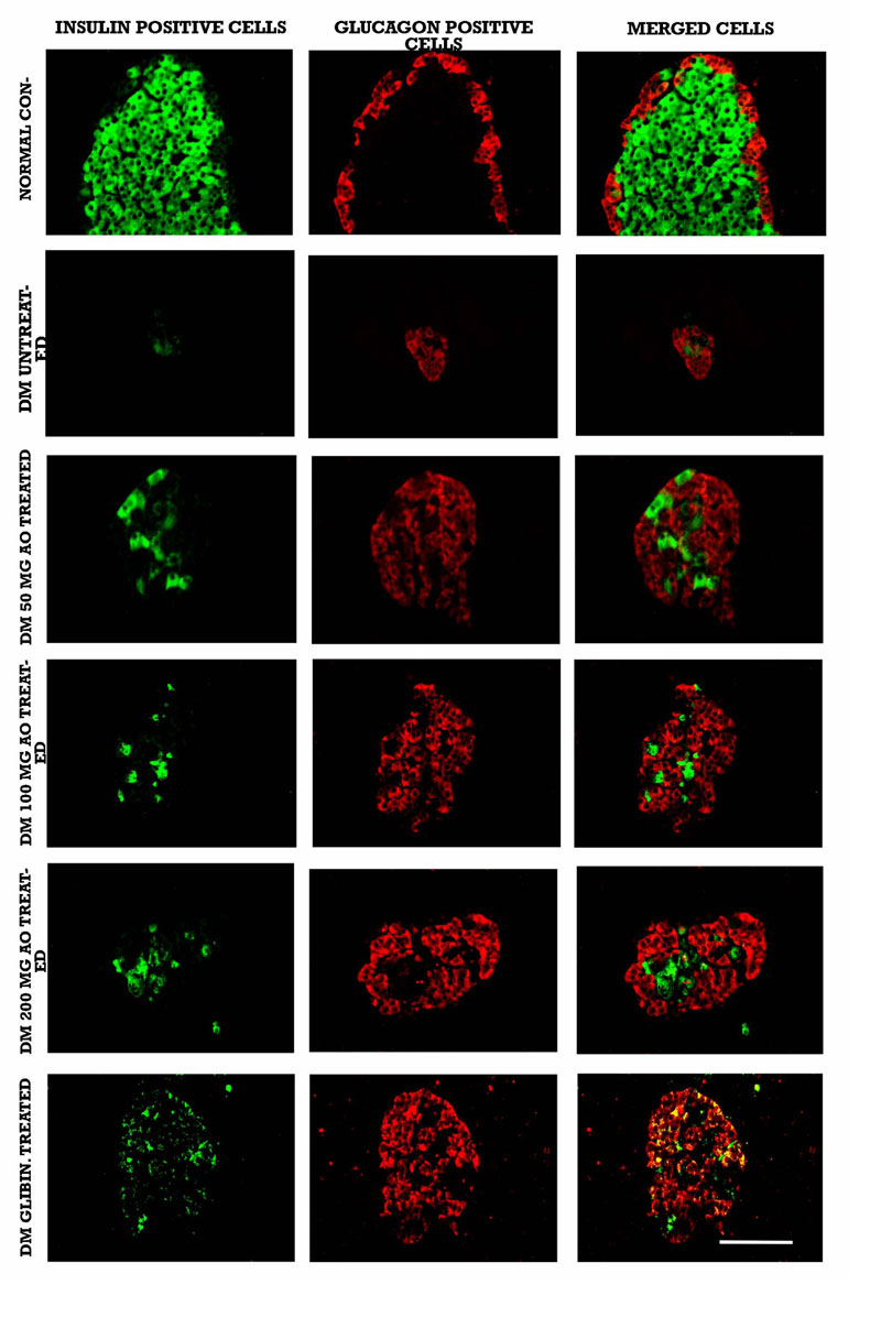
Fig. (2) Immunofluorescence double-labelling images depicting insulin-immunoreactive cells (green) and glucagon-positive cells (red) in the islets of normal, untreated diabetic, and Acridocarpus orientalis (AO)-treated diabetic rats. Note the increase in the number of insulin-positive cells in the islets of AO-treated diabetic compared to untreated diabetic rats. n=6 for each data set. Scale bar = 25 µm.