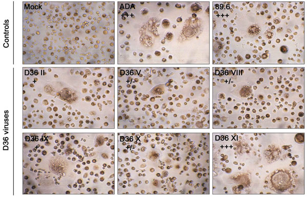
Fig. (3). Syncytium formation in MDM induced by nef-deleted viruses MDM were infected with equivalent amounts of each virus, as described in Materials and Methodology. Mock-infected MDM were treated with culture medium alone. Syncytia formation was documented at day 11 (ADA), 14 (89.6) or 18 (D36 viruses) post-infection. Syncytia were counted manually and scored as +/-, occasional syncytia; +, low frequency of syncytia, occurring in <5% of cells; ++, moderate frequency of syncytia, occurring in 5 to 50% of cells; or +++, extensive syncytia, occurring in >50% of cells, as described previously [37]. Results are representative of two independent experiments using cells obtained from different donors, which gave similar results. Photographs are at a final magnification of x400.