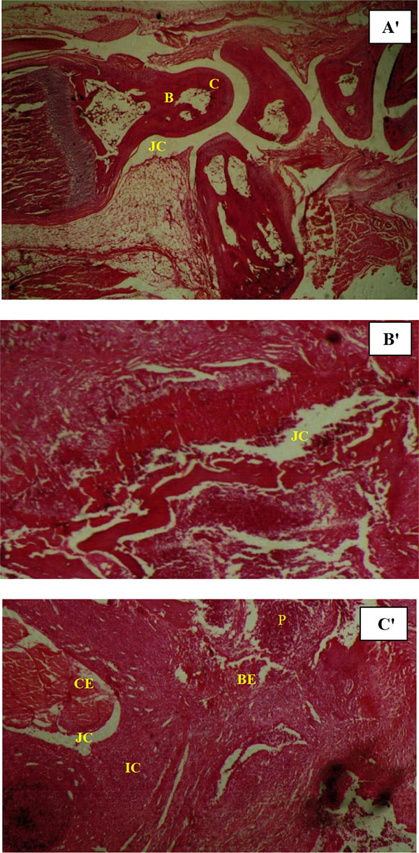
Fig. (6)
Hematoxylin-eosin stain preparation of organ sections. Histopathological analyses in a hind paw of normal rat (A'); rat model of septic arthritis (B'); and leg joint in a treatment group (T2) rat (C'), C: cartilage; B: bone; BE: bone erosion; CE: cartilage erosion; JC: joint cavity; IC: inflammatory cells and P: pannus formation. Histopathological micrographics are shown with 10x magnification.