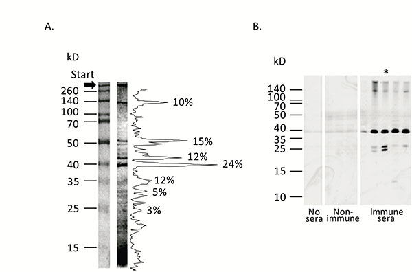
Fig. (2)
Analysis of staphylococcal anatoxin (crude cell-free liquid cultures of S. aureus).Panel A. Protein composition of staphylococcal anatoxin. Staphylococcal anatoxin was run on 10% SDS-containing polyacrylamide gel and stained with silver nitrate [23]. Protein mass markers are shown on the left. Quantification of the protein bands was accomplished with ImageJ program [54] and shown on the right. Proportion of the most intensive bands is shown as percentages.
Panel B. Immunogenic properties of staphylococcal anatoxin. Staphylococcal anatoxin was subjected to Western blot. Afterwards nitrocellulose membrane was left untreated (“No sera”, two tracks), treated with mouse sera obtained from non-immunized animals (“Non-immune”, three tracks) or treated with mouse sera obtained from immunized by absorbed staphylococcal anatoxin animals (“Immune sera”, 5 tracks). Each track represents reaction with an individual serum. The membranes were incubated with anti-mouse peroxidase conjugate and developed with chemiluminescence reagents. Approximate positions of molecular mass markers are shown on the left. Asterisk indicates the serum, used in subsequent experiments to identify hyper immunogenic staphylococcal components.