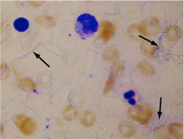
Fig. (2).Borrelia spirochetes concentrated from 10 ml blood and Giemsa stained. Low numbers of bacteria were concentrated as described in the material and methods section, Giemsa stained and visualized by light microscopy at 1000X magnification. Cells stained red and blue are contaminating erythrocytes and leukocytes, respectively. Some of the bacteria are marked by arrows.