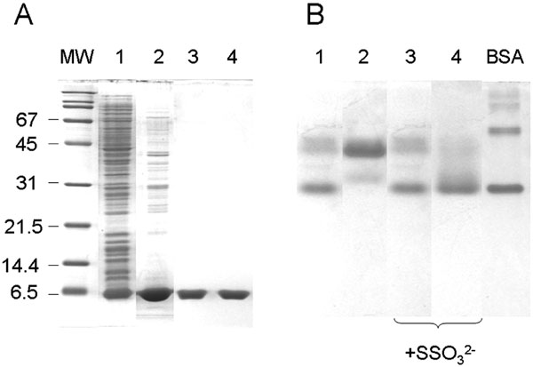
Fig. (3) Electrophoretic analysis of PspE1 and PspE2. A: Fractions obtained during purification of PspE from induced strain BL21(DE3)(pHC4.1) were analyzed on an SDS-polyacrylamide gel. Lane 1, total cellular proteins (10 μg). Lane 2, freeze-thaw extract (10 μg). Lanes 3 and 4, pooled peak 1 (PspE1) and peak 2 (PspE2), respectively, from cation-exchange chromatography (3 μg each lane). MW, molecular weight standards. B. PspE1 (lanes 1 and 3) and PspE2 (lanes 2 and 4) were analyzed using nondenaturing polyacrylamide gel electrophoresis. In each lane, 3 μg of PspE was analyzed. Where indicated, PspE was pre-incubated with 1 mM ammonium thiosulfate for 5 min at 22oC. BSA, 2 μg BSA (monomeric and dimeric forms are the two major bands) was used as a standard.