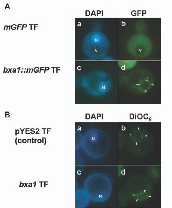
Fig. (4) Localization of Bxa1::mGFP fusion protein in yeast transformants. (A) Green fluorescent image derived from Bxa1::mGFP are shown by arrows in d. Nucleus [N] in yeast cells were visualized by DAPI staining in a and c. Vacuole [V] was detected as a dark area without any fluorescent signal in a and b. (B) Double staining of pYES2 transformant (control strain) and bxa1 transformant by DiOC6 and DAPI. Green fluorescent image derived from DiOC6 is indicated by white arrow heads in b and d. Nucleus [N] in yeast cells were visualized by DAPI staining in a and c.