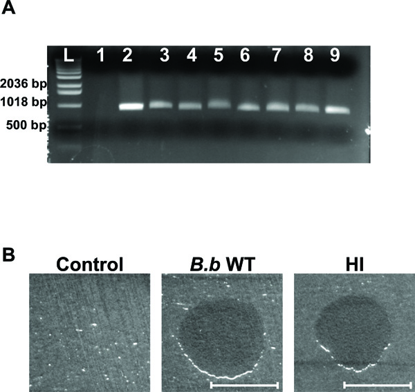
Fig. (2) HI variant analysis. (A) Agarose gel electrophoresis of amplified DNA with B. bacteriovorus specific primers. Lanes correspond to template E. coli strain WM3064 DNA (lane 1), template B. bacteriovorus 109J WT DNA (lane 2), template DNA from HI variants (lanes 3-9), and 1 Kb DNA ladder (Invitrogen, Carlsbad, CA) (L). A ~900 bp fragment was obtained for all of the 35 HI variants examined, with seven random HI variants shown here. (B) Plaque predation assay. Wild-Type B. bacteriovorus (B.b WT), HI variant (HI) and DNB (Control) were spotted on a thick lawn of Klebsiella pneumoniae host cells. 48 hr after inoculation a clear lytic halo formed at the point of inoculation where predation had occurred. Lytic halos appeared for all of the 35 HI isolates examined. Scale bar, 0.8 cm.