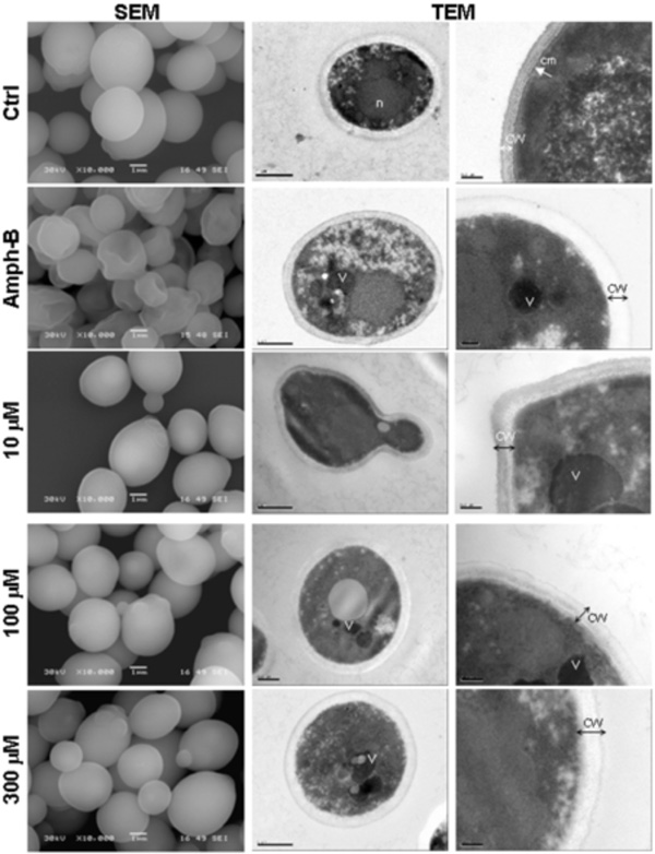
Fig. (2) Scanning electron microscopy (left column) and transmission electron microscopy (two right columns) micrographs of C.
albicans with and without farnesol.Candida was untreated (Ctrl), treated with Ampho-B (positive control) or with farnesol at various concentrations
for 24 h then subjected to scanning electron microscopy (SEM) and transmission electron microscopy (TEM) analyses. (n), nucleus;
(cm), continuous cytoplasmic membrane; (CW), Cell wall; (V), vacuole. Each experiment was repeated three and four times, for SEM
and TEM, respectively.