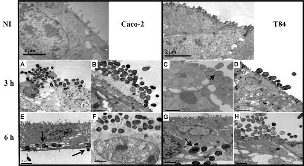
Fig. (3) Transmission electron microscopy of differentiated Caco-2 (A, B, E and F) or T84 (C, D, G and H) cells fixed at 3 h or 6 h after infection with aEPEC 1711-4 (A, C, E and G) or aEPEC 3991-1 (B, D, F and H). Note the formation of large bacterial clusters and pedestal formation at the apical surfaces of cells at 3 h after infection. Partial internalization with presence of electron dense material below bacteria (C – black arrow head) indicating actin accumulation can be detected at 3 h after infection with aEPEC 1711-4. Black arrows indicate aEPEC 1711-4 residing within vacuoles and forming pedestals at the basolateral portion of enterocytes (E and G). NI represent uninfected Caco-2 and T84 cells respectively.