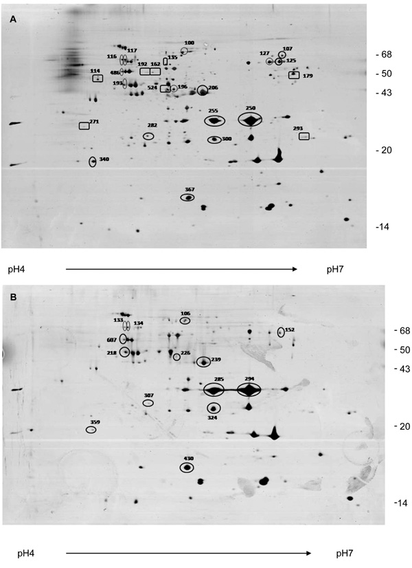
Fig. (3). Two-dimensional electrophoresis pattern of OMP extracts from aEPEC (A) and tEPEC strains (B) grown in LB broth. OMP extracts were applied to 4–7 IPG strips for isolectrofocusing followed by electrophoresis on 12% polyacrylamide gel. The gels were silver-stained. Spots indicated with numbers were identified by in-gel trypsin digestion and MALDI-TOF MS. Circle: proteins identified in both strains. Square: proteins identified only in aEPEC.