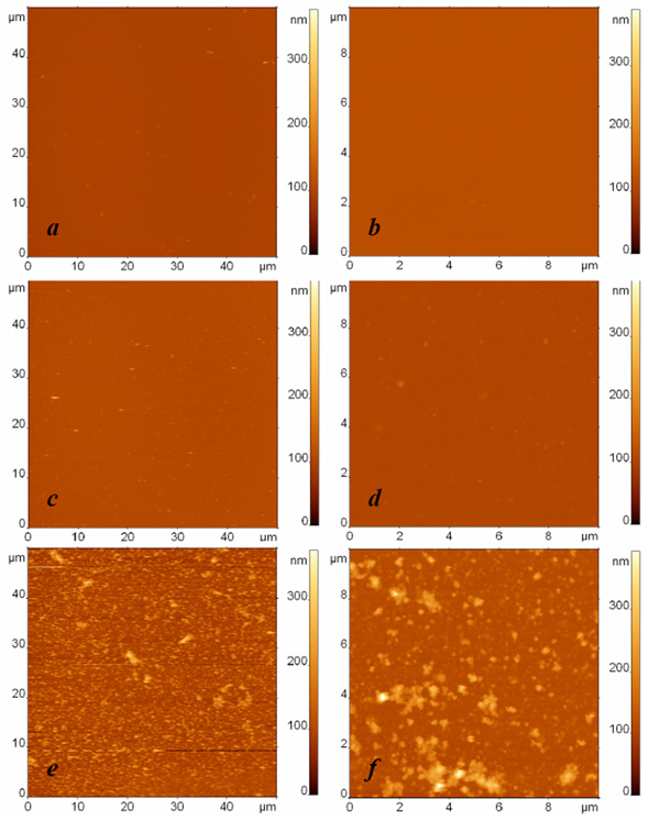
Fig. (2) AFM images of surfaces modified with protein G and anti-Escherichia coli antibodies and exposed to (a, b) the buffer, (c, d) the
suspension containing Bacillus subtilis cell fragments with the concentration 108 cells/ml, and (e, f) the suspension containing Escherichia
coli cell fragments with the concentration 108 cells/ml. The surfaces were rinsed by immersion in the buffer and rotation in the shaker. The
images sizes are (a, c, e) 50 µm and (b, d, f) 10 µm. The height difference is 400 nm in all images.

