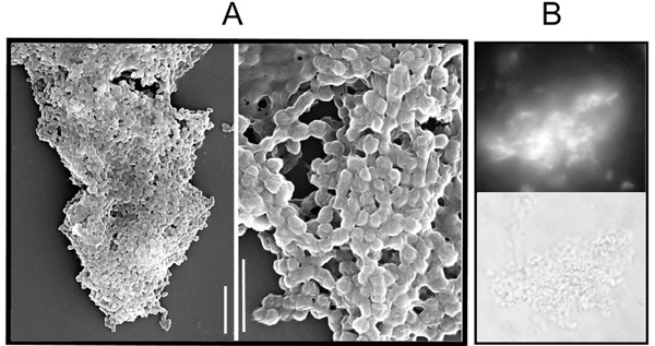
Fig. (2) Microscopic observations of B. abortus aggregation under microaerobic conditions. (A) Scanning electron micrograph of B. abortus 2308; scale bars: 5 µm (left) and 2 µm (right). (B) Fluorescence (upper) and phase-contrast (lower) micrographs of B. abortus vjbR stained with calcofluor. X 100.