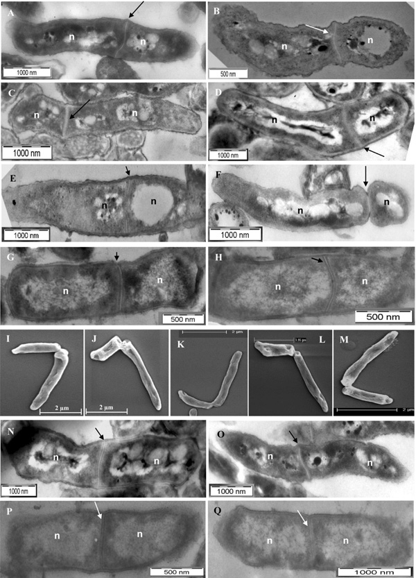
Fig. (1)
TEM and SEM imaging of mid-log phase M. smegmatis and M. xenopi
cells with highly deviated asymmetric septum/ constriction or symmetric septum.
(A-D) TEM images of M. smegmatis cells with highly deviated
asymmetric septum. (D) M. smegmatis cell undergoing ‘snapping
post-fission’ mode of highly deviated asymmetric division. (E)
M. smegmatis cell at the initiation of the septum formation. (F)M. smegmatis cell close to the completion of the asymmetric septum
constriction. (G, H)M. xenopi cells with highly deviated
asymmetric septum. (I-M)SEM images of M. smegmatis cells
with ‘snapping post-fission’ mode of highly deviated asymmetric division.
(N, O)M. smegmatis cells with symmetric septum with minor
deviation. (P, Q)M. xenopi cells with symmetric septum
with minor deviation. Arrow indicates the position of the highly deviated
asymmetric septum or constriction, or symmetric septum with minor deviation, in
the septum position from the mid-cell site (see Table 1). n indicates
nucleoid.