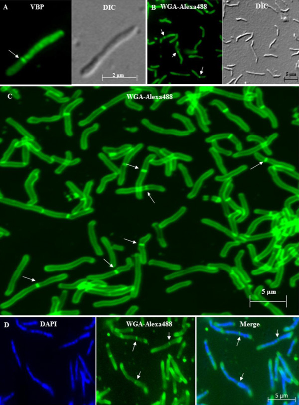
Fig. (3)
Fluorescence imaging of mid-log phase live and fixed M. smegmatis and
M. xenopi cells with asymmetric septum.
(A).
Fluorescence and the corresponding DIC image of the VBP stained live M.
smegmatis cell with highly deviated asymmetric septum.
(B). Fluorescence and the corresponding DIC image of the WGA-Alexa488
stained fixed M. smegmatis cell with highly deviated asymmetric septum.
(C). Confocal image of fixed WGA-Alexa488 stained M. smegmatis
cells with highly deviated asymmetric septum. (D). Highly deviated,
asymmetrically dividing M. smegmatis cells stained with DAPI for nucleoid
and WGA-Alexa488 for septum. The merge figure shows the WGA-Alexa488 stained
septum dividing the DAPI stained nucleoids. In all the panels, the arrows
indicate the position of the highly deviated asymmetric septum.