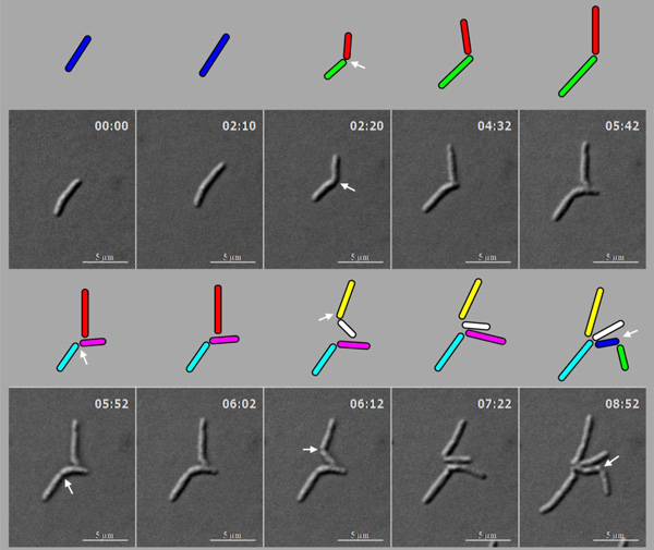
Fig. (4)
Live cell time-lapse imaging of highly deviated asymmetric division of M.
smegmatis cells, with colour cartoon for the cell images.
Only minimum number of panels are shown just enough to depict the phenomenon. An
M. smegmatis mother cell (blue) first underwent symmetric division to
generate daughter cells of lengths, 3.08 µm (red) and 3.20 µm (green), with 0.12
µm difference in their lengths. One of the daughter cells (red) further
underwent asymmetric division, generating a short daughter cell (2.28 µm; white)
and a long daughter cell (3.66 µm; yellow), with 1.38 µm difference in their
lengths. The other daughter cell (green) underwent symmetric division,
generating daughter cells of lengths, 4.06 µm (pink) and 4.26 µm (cyan), with
0.2 µm difference between their lengths. One of these daughter cells (pink cell;
4.06 µm) further grew and underwent symmetric division to generate two daughter
cells of lengths, 2.60 µm (blue) and
2.87 µm (green). Thus, the daughter cells of a symmetric division underwent two
different modes of division. The growth rate of the short and the long cells of
the asymmetric division were determined from the live cell imaging panels.