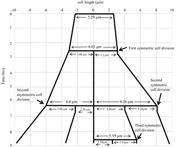
Fig. (5)
The lineage of the growth and highly deviated asymmetric division of live M.
smegmatis cell and of its daughter cells, shown in Fig. (4).
The growth and division lineage was traced from the images of time-lapse
microscopy in Fig. (4). The zero time point does not correlate with the
birth of the starting mother cell.