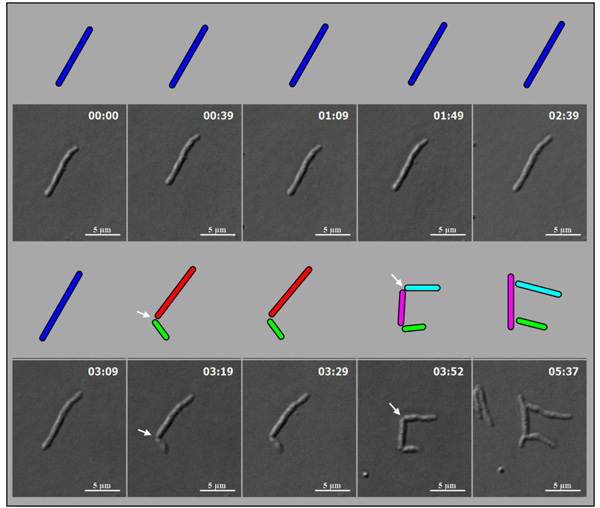
Fig. (6)
Live cell time-lapse
imaging of highly deviated asymmetric division of mid-log phase
M. smegmatis long mother cell.
An M. smegmatis long mother cell (blue cell; 9.79 µm) first
underwent highly deviated asymmetric division close to one pole
(arrow), to generate a short daughter cell (green cell, 2.67 µm) and
a longer-sized daughter cell (red cell, 7.84 µm), with the
difference of 5.17 µm in their lengths. The longer-sized daughter
cell (red cell, 7.84 µm) subsequently showed symmetric division with
minor deviation (arrow), generating daughter cells of lengths, 4.59
µm and 4.36 µm (pink and cyan cells), with the difference of 0.23 µm
in length between them. The images were observed under DIC. Arrows
indicate the site of cell constriction at asymmetric or symmetric
position.