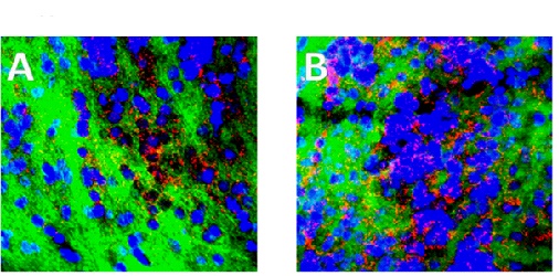
Fig. (3)
LPS treatment resulted in a substantial, sustained increase in inducible nitric oxide synthase (iNOS) for at least 12 hours Fig. (3) shows images from sections of olfactory bulb at 2 and 8 hours post treatment), with iNOS (Red) clearly visible concentrated along the nuclei (blue) of sensory cell tracts. The connective tissue matrix is shown in green (Magnification x 900). Cortical tissue was used as “control” as our initial experiments concentrated on the olfactory bulb as loss of olfaction is an early indicator of PD, but when comparing cortex and olfactory bulb there was a substantial increase in iNOS in both areas, attesting to a major and prolonged inflammatory response, but these images not shown.