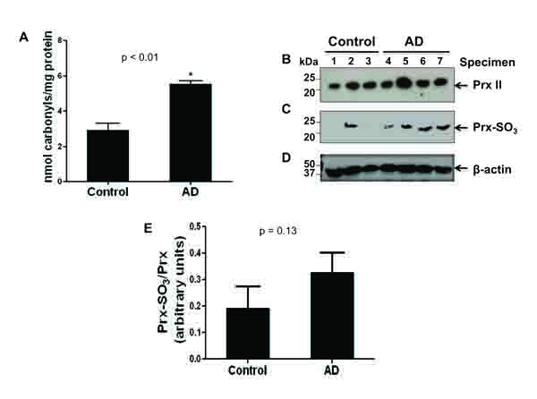
Fig. (1)
Presence of increased amounts of carbonylated protein, Prx II and its inactive Prx isoform in tissue samples from AD brains. (A) The amount of protein carbonylation in cytosolic fractions from the frontal cortex of AD and control brain specimen was measured by ELISA as described in the Method Section. Protein (1 μg) from each of the 7 samples (AD, n=4; control, n=3) was affixed to a 96-well assay plate, and probed for carbonylation via an antibody against DNP. A graph of the amount of carbonyls (nmol carbonyls) per mg of protein is shown. *p<0.01, significantly different from the control samples. (B-D) Cytosolic proteins of both AD and control samples (100 μg/well) were separated on 15% SDS-PAGE, transferred to PVDF-Immobilon membranes, and probed with the specific antibodies against Prx II (B), Prx-SO3 (C), or β-actin (D), used as a loading control. (E) The densitometric quantitation of the immunoblots in B with Prx-SO3 normalized to Prx II is presented.